- If you only need to check the
Cameca's spectrometers, or if you are just starting with
a list of elements in hand, you can now begin considering
the configuration for the microprobe.
- Because a single x-ray
spectrometer may have several diffracting crystals from
which to choose, many possible configurations for the
Cameca exist. The TV monitor will indicate the
Cameca's current crystal configuration ... but our
Cameca's spectrometers also offer the following
possibilities ...
| spectrometer
1 |
spectrometer
2 |
spectrometer
3 |
spectrometer
4 |
soft
x-ray
(low pressure) |
soft
x-ray
(low pressure) |
hard
x-ray
(high pressure) |
hard
x-ray
(high pressure) |
TAP
lead sterate
PC1
PC3 (soon) |
TAP
PET |
PET
LIF |
PET
LIF |
- ... if you need to
change a crystal use the Probe for Windows software or be
sure to move to a spectrometer position greater than
81000 first. The SX commands are:
-
- SX> AMOV spN "position"
(e.g., AMOV SP2 81000)
- SX> XTAL spN "xtal
name" (e.g., XTAL SP2
PET)
-
The two most common
configurations for the spectrometers are:
| spectrometer |
SP1 |
SP2 |
SP3 |
SP4 |
| most
minerals |
TAP |
PET |
LIF |
LIF |
| feldspars |
TAP |
TAP |
PET |
LIF |
- The first allows spectrometers
3 & 4 to share several transition metal elements
which are typically counted for longer periods of time.
The second is optimized for a mineral like
feldspar which involves few metals but allows
spectrometers 1 & 2 to share Na, Al, Si and allows Mg
and Fe free of charge. Notice how these two
possible configurations ask you to organize your work
over two different sessions, but your time on the
instrument will be less overall.
| crystal |
PC1 |
OdPb |
TAP |
PET |
LiF |
wavelength
range (A) |
51 to 12 |
85 to 20 |
22 to 5.2 |
7.35 to 1.75 |
3.42 to 0.81 |
| K
lines |
C(6)
to F(9) |
B(5)
to O(8) |
F(9)
to P(15) |
Si(14)
to Cr(24) |
Sc(21)
to Br(35) |
| L
lines |
n.a. |
Ca(20)
to V(23) |
Mn(25)
to Y(39) |
Sr(38)
to Eu(63) |
Te(52)
to Bi(83) |
| M
lines |
n.a. |
n.a. |
La(57)
to Ir(77) |
W(74)
to Pu(94) |
n.a. |
- The table above and Cameca's sine-theta
table imply you
have some choices ... for some elements you can either
measure an element's specific x-ray line with a choice of
crystals or you may have a choice of measuring it K, L or
M line. A general rule of thumb is to pick the
line with the best signal-to-noise ratio ... another
generality is that best S:N ratios exist for K lines and
locations on any crystal for large sine-theta values.
An exception for picking the best S:N crystal is
for major elements when the best S:N isn't needed and
because spectrometer reproducibility is better for small
sine-theta values. For example, this would imply
you would choose TAP for Si in silicates but would choose
PET for Si in spinels.
- Regarding your choices between
K,L or M lines, choose the best S:N ratio when needed for
sensitivity, but also concern yourself with spreading the
elements around. For example, instead of measuring
Cu K in sulfides with LiF measure the L line with the
idle TAP spectrometer.
Pulse Height Analysis
- PHA can be thought of as
"Pulse Height Acceptance" After
x-rays enter the detector and are amplified the PHA does
not allow the x-ray counter to count unwanted pulses.
The PHA (1) typically excludes noise by setting a baseline,
and (2) the EPMA analyst may also choose not to count
higher energy pulses by setting a window or an upper
threshhold. The Cameca's mode of PHA detection
can be set to use only a baseline (integral) or to use a
window (differential).
SX>
SACQ spN MODE INTE (count
all pulses above the baseline voltage)
SX> SACQ spN MODE DIFF
(count only pulses within the window)
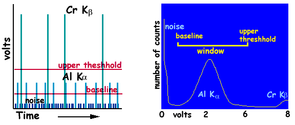
- A good example of PHA and its
benefits is depicted by the measurement of Al x-rays
while chromium is present ... e.g., in Cr-spinel.
That is, the Cr K-beta line's wavelength is the 4th order
equivelent of Al K-alpha ... the primary difference being
its energy ... four times that of the Al line.
Therefore ... as shown below ... the upper threshhold can
exclude Cr x-rays from Al's measurement.
- The optimum window settings
can be set by the Cameca by issuing the command:
SX> ADJP spN or
SX> ADJP SPEC
(all spectrometers)
... which tells the Cameca
to have the window follow the spectrometer's position.
We have found this to be accurate and more
advisable than determining what is optimum yourself
because it is consistent with what everyone else uses.
- The window width for the
differential mode is generally a consideration for which
crystal is used. You can set the window width with
the following command:
SX>
ADJP spN CW X (where X= 1,2,3,4,5)
- The following table indicates
general guidelines for the CW parameter:
| crystal |
PC1 |
OdPb |
TAP |
PET |
LiF |
| Cw |
2 |
2 |
3 |
5 |
5 |
- The PHA is sensitive to the detector gas
and the HV bias applied to the detector. You don't
have much control over the gas pressure, but you do over
the bias ... it is the primary parameter for making sure
the PHA is adjusted properly. You first have to
test the PHAs:
(1)
Move the stage to a standard which can be a source
for all x-rays:
SX> MOVE STAG ANDR
(move stage to andradite garnet)
(2) Move the
spectrometers to the element x-ray positions
SX> MOVE SP1 SI KA
(move TAP crystal to Si x-ray position)
SX> MOVE SP2 CA KA
(move PET crystal to Ca x-ray position)
SX> MOVE SP3 FE KA
(move LiF crystal to Fe x-ray position)
SX> MOVE SP4 FE KA
(move LiF crystal to Fe x-ray position)
(2) Test the PHAs
SX> TPHA SPn
(test each one at a time ... adjust bias if needed)
- Adjusting the bias will center
the x-ray counts within the window (... baseline and
threshhold ...). Note the current BIAS setting and
you can shift the peak left or right by modifying the
voltage by as little as 5 volts (... try 10 volts at
first ...).
- You can set the BIAS with the
command:
SX> SACQ SPn
BIAS XXXX (where XXXX is the bias voltage
value)
Verifying the
Spectrometers
- The Probe for Windows software
maintains the spectrometer offsets internally using a
polynomial fit to calibrated positions separately for the
K, L and M x-ray line families. This is preferred to the
Cameca "veri spec" command assumes a constant
offset over the entire range of the spectormeter.
-